Medical product development

First product:
DermaFC

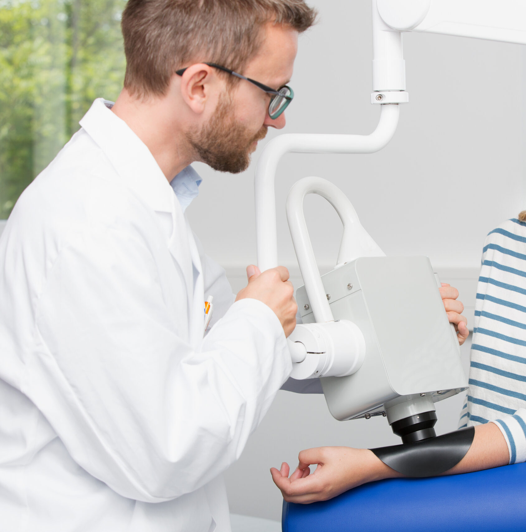
Dermatofluoroscopy is a method for the early detection of malignant melanoma.

This patented technology is based on the selective excitation of melanin fluorescence in the skin. The skin lesion suspicious of malignant melanoma is scanned with the laser and Magnosco’s machine learning algorithm analyzes the data and provides a score that guides the physician to a reliable diagnosis. The Magnosco DermaFC, in which the method has applications, is in use for study purposes only.

Dermatofluoroscopy

Under normal excitation conditions, the ultra-weak melanin fluorescence is completely outshone by the autofluorescence of other endogenous fluorophores in the skin such as NADH, tryptophan, flavin, or lipofuscin. Using stepwise two-photon absorption, melanin can be selectively excited and its fluorescence becomes visible.

In dermatofluoroscopy, melanocytes of healthy skin, melanocytes of nevi, and melanocytes of melanomas show characteristic fluorescence spectra that allow reliable differentiation between benign and malignant tissue.

For the excitation of melanin by stepwise two-photon absorption, nanosecond laser pulses are focused at 800 nm in the epidermis. The resulting melanin fluorescence is detected in the range of approximately 385 – 785 nm. Using this excitation technique, the lesion is scanned point by point.

Do you have any questions?
Here, we answer the most important things for you.

Commercial use of the DermaFC on the German market is currently not possible, as we are dedicated to the further development of dermatofluoroscopy and other medical products.

The calibration and validation of the DermaFC were carried out as part of a three-year, multicenter clinical trial at the University Hospital of Tübingen, the University Hospital of Heidelberg, and the Charité University Hospital Berlin. We are constantly conducting further studies to verify the results as well as to further develop the method.

Magnosco DermaFC combines a patented, physical measurement method and AI. The underlying method (dermatofluoroscopy) uses a novel approach in which the patented technology enables specific excitation of melanin fluorescence in the skin. The complex evaluation of the recorded spectra is performed by AI-based data analysis.

We are continuously developing the method to support precise excisions, histological examinations, and rapid diagnosis of other skin cancers. First investigations show fields of application in other skin cancers, inflammatory lesions, or also in cosmetics.